
Hematology of Leukemia
Overview
The Woburn Toxic Trial focused on a suspected childhood leukemia cancer cluster and its connection to pollutants in the municipal water supply. This webpage includes background information on the hematology of leukemia.
Components of Human Blood
Human blood is an amazing fluid. It consists of plasma and cells (red blood cells, white blood cells, and platelets) that supply nutrients and other elements to tissues and remove waste products. Blood also enables other cells and substances to be transported between tissues and organs, transfers heat to the skin, and acts as a buffer to regulate pH. About five liters of blood circulate in an average adult. Blood is created solely by the marrow inside bones.

Red blood cells are the most common type of blood cell and are the body's principal means of delivering oxygen from the lungs to tissues. White blood cells (also known as leukocytes) help the body defend against infectious disease and foreign materials as part of its immune system. Platelets cause blood to clot.
Leukemia -- Cancer of the Blood
Leukemia is a disease of the white blood cells. A leukemia patient has an elevated white blood cell count consisting largely of immature and unhealthy white blood cells. Normal bone marrow cells are displaced by increasing numbers of the malignant cells, which results in a lack of blood platelets and regular white blood cells needed to clot blood and fight infection.
Leukemia is categorized as either acute or chronic. In acute leukemia, immature blood cells rapidly grow and the bone marrow is unable to produce healthy blood cells. In chronic leukemia, there is an excessive buildup of relatively mature, but abnormal, blood cells. Chronic leukemias typically take months or years to progress. Leukemia is also classified by the type of abnormal blood cell found most often. When leukemia affects lymphoid cells, it is considered lymphocytic leukemia. When myeloid cells are affected, it is considered myelgenous leukemia.
Combining these two types of categories, leukemia is further categorized as:
- Acute lymphocytic leukemia (ALL), which is also known as acute lymphoblastic leukemia). ALL is the most common type of leukemia found in children. The disease also affects adults, particularly those over 65 years of age.
- Acute mylogenous leukemia (AML) is more common in adults than in children.
- Chronic lymphocytic leukemia (CLL) occurs most often in adults over the age of 55.
- Chronic myelogenous leukemia (CML) occurs in adults, with a very small number of children developing this disease.
The causes of leukemia are unknown but genetic and environmental factors are involved. Some studies link leukemia to exposure to high doses of radiation and petrochemicals. Higher leukemia rates are found in more developed countries and in higher socioeconomic groups.
Acute Lymphocytic Leukemia
ALL is the type of leukemia most often found in children under the age of 19. About 3,970 cases are diagnosed each year in the United States. ALL results from an injury to the DNA of a cell in the bone marrow. A leukemic cell replaces normal bone marrow, which leads to:
- Uncontrolled and exaggerated growth and accumulation of cells ("lymphoblasts" or "leukemic blasts". These cells don't function as normal blood cells.
- Blockage of normal bone marrow cells, leading to a deficiency of red cells (anemia), platelets (thrombocytopenia), and normal white cells (especially neutrophils) in the blood.
- Possible symptoms of childhood ALL include: fever, easy bruising or bleeding, petechiae (flat, pinpoint spots under the skin caused by bleeding), bone or joint pain, painless lumps in the neck, underarm, stomach, or groin, pain or feeling of fullness below the ribs, weakness or feeling tired, and loss of appetite.
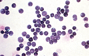
Bone Marrow


The two images at left are from needle aspiration biopsies of human bone marrow, a medical procedure that pierces the bone marrow with a thin needle to collect cells for examination. The cells are then smeared on a slide for examination under a microscope. The image on the left shows normal bone marrow cells and a moderate number of lymphocytes (the purple-stained cells) distributed throughout the smear. In the blood smear of an ALL patient, as seen at right, lymphoblasts and undeveloped lymphocytes dominate and are present at unusually high concentrations. Lymphoblasts, which give their name to acute lymphoblastic leukemia, are lymphocytes that have enlarged to fight off antigens. In leukemic marrow, however, malignant lymphoblasts reproduce uncontrollably, even in the absence of antigens.


The photographs of marrow at right show marrow as it appears in the human body. In normal bone marrow, seen center right, many different cell types are present, and the large, white blood cells are distributed in great quantity throughout the marrow. The different cell types in marrow can be seen at center in an illustration of bone marrow. In the marrow of an ALL patient, far right, the cells are very dense and lymphoblasts dominate. This is especially apparent when you examine the small number of white blood cells that remain. The consequences of lymphoblast growth is that the stem cells needed to produce red blood cells and healthy white blood cells are squeezed out of the marrow, with the cancerous cells consuming their nutrients and space.
Blood


Cerebrospinal Fluid
Trial Issues and Leukemia Treatment
The plaintiffs alleged a causal connection between prolonged consumption of the contaminated drinking water and the victims of leukemia. They relied on Harvard medical studies and the Massachusetts Department of Health as primary sources of information regarding the relation between the chlorinated solvents, TCE and PCE, and the cancers. The defendants utilized experts to discredit the accuracy of the Harvard and Massachusetts studies. The defendants used medical science studies showing the connection between "every day" items and related high incidence of cancers. Their approach focused more on creating a reasonable doubt between cancers and its causes.Mock Trial Strategies and Leukemia Treatment
Students should be encouraged in their preparation for the mock trial to research the scientific evidence connecting leukemia to specific sources. They should look at improvements in treatment and in understanding the mutation that results from exposure to certain environmental conditions. Over the past decade, our understanding of the relation between the cause-and-effect of certain cancers has dramatically improved.